Enhanced Cortical Inhibition and Reduced Sound Responsiveness in Ossicular Disruption (OD) Rats
Do Eun Kim1, Jong Chan Jeon1, Bohyeon Park1, Nahae Park1, So Young Kim1,2
1Department of Anatomy and Cell Biology, Seoul National University College of Medicine, Seoul, Republic of Korea
2Sensory Organ Research Institute, Seoul National University Medical Research Center, Seoul, Republic of Korea
Background: Conductive hearing loss reduces auditory input without directly damaging cochlear or auditory nerve structures, providing a model to examine how diminished sensory drive affects central auditory and limbic circuits. However, how chronic auditory attenuation alters sound-evoked activation in higher brain regions remains unclear.
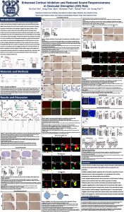
Methods: We used an ossicular disruption (OD) rat model to chronically reduce auditory input while preserving early brainstem responses. Sound-evoked neural activity was assessed using c-Fos immunohistochemistry and in situ hybridization following sound exposure either in the home cage (passive) or during maze-like open field exploration (MOF), allowing evaluation of behavioral context–dependent modulation. To examine changes in sound-responsive cellular populations, we performed cell-type–specific and multiplex immunofluorescence targeting parvalbumin (PV) interneurons and Iba1-positive microglia. Open field and Y-maze tests were conducted under baseline and sound conditions to assess exploratory behavior and locomotion.
Results: Ossicular disruption significantly elevated auditory brainstem response thresholds, confirming sustained auditory attenuation. Under passive sound exposure, c-Fos expression was markedly reduced in the auditory cortex (CTRL: 967.8 vs. OD: 324.7 cells/mm², p < 0.0001), with no change in somatosensory cortex. In contrast, during MOF exploration, sound-evoked c-Fos expression in the auditory cortex and amygdala was significantly increased in OD animals compared with controls. This context-dependent recruitment was accompanied by increased PV interneuron density and enhanced c-Fos overlap with PV interneurons and Iba1-positive microglia. Behaviorally, OD animals showed increased center exploration and locomotor engagement during sound exposure without cognitive deficits.
Conclusion: Chronic conductive hearing loss reorganizes auditory and limbic circuit activation in a context-dependent manner, suppressing responses during passive listening while enhancing engagement during active exploration. These changes involve altered inhibitory circuitry and cellular recruitment, indicating reshaped sensory–behavior integration rather than uniform central suppression.